Confocal microscopy – illinois state university advanced bioimaging 2: schematic of a basic confocal microscope. 1: schematic representation of a confocal microscope (extracted from a
Confocal Microscopy – Illinois State University Advanced Bioimaging
Laser scanning confocal microscope diagram
Confocal microscopy microscope
*extended topic* microscopy enhanced by the wave characteristics ofSchematic image of the operating principle of a confocal microscope Confocal tutorial: priniples: what is confocal? amu dept pathol univ helConfocal microscope- definition, principle, parts, types, labeled.
Confocal microscope schematic builtConfocal microscope microscopy schematic thorlabs pinhole system upgrade wheel motorized Confocal microscopyConfocal imaging.

Confocal microscopy principle ppt kelly david november conjugate pinhole powerpoint presentation
Confocal microscopyZeiss microscopy online campus Confocal laser scanning microscopy in 3 simple stepsFigure b.1: schematic diagram of a conventional confocal microscope.
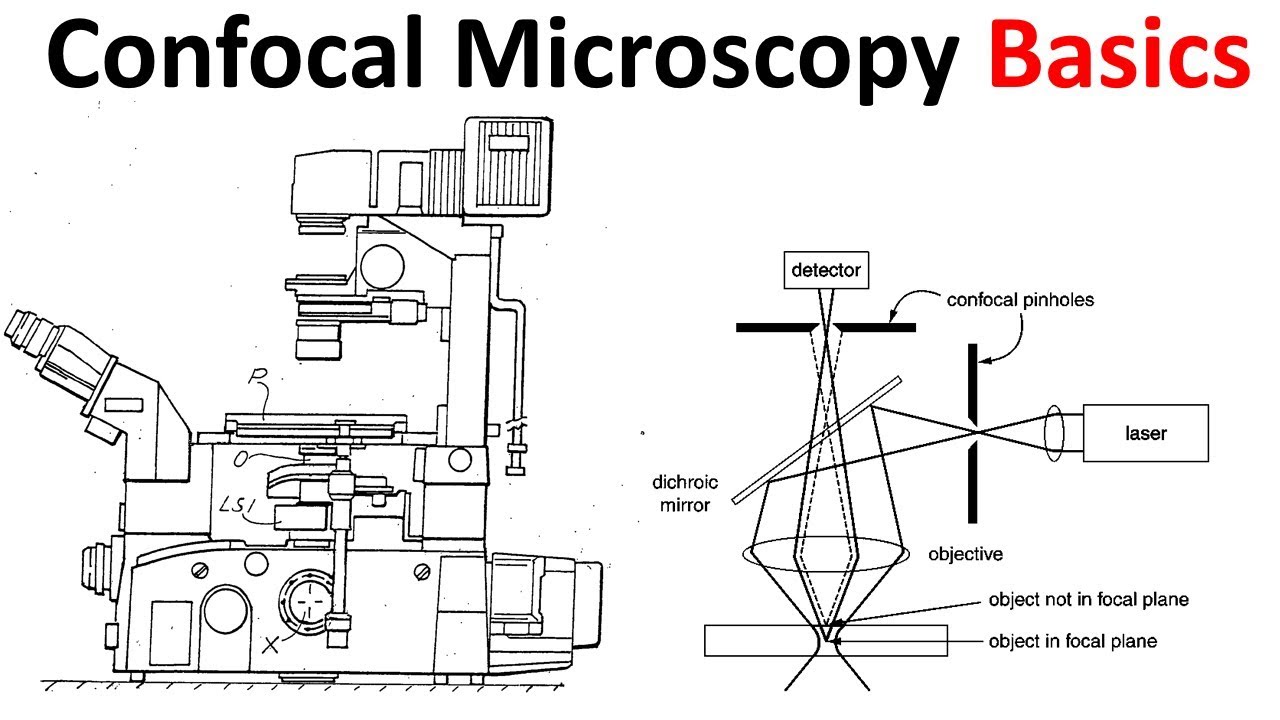
Confocal microscopesConfocal pinhole microscopy schematic detector Confocal microscopy basicsSchematic diagram of a confocal microscope for measuring surface.

Confocal microscope microscopy laser gif formation amu technology illumination project tutorial cf
Confocal light microscopy microscope path vs plane extended beam physics focus focal laser pinhole schematic field characteristics wave photons chapterSchematic diagram of a confocal microscopy. the detector pinhole, point Confocal microscopy microscope light schematic pinhole widefield below left rightHigh resolution confocal laser scanning microscopy.
Confocal optical path thorlabs microscope scanning laser microscopy imaging figure cerna channel four systems based tutorialSchematic diagram of the confocal microscope. Confocal microscopy microscope principleConfocal microscope leica sp8 path microscopy bioimaging facility imaging multichannel illinoisstate.

Confocal microscopy applications
Confocal microscope principle schematic operating straigth11: schematic depiction of a typical confocal microscope. The benefits of confocal microscopy in dermatology – eadvvienna2020.orgMicroscope confocal microscopes britannica microscopy encyclopædia.
Confocal microscope measuring reprinted topographyConfocal imaging Confocal microscope principle pinhole light focus imaging fluorescent objective collimated depth excitation focal focused beam pointConfocal microscopy – working principle and applications in dermatology.
Confocal microscopy
Microscope confocal schematic obtain spectra fluorescence(a) schematic of the custom-built confocal microscope; (b) 3d view of Confocal microscopy upgradeConfocal microscopy.
Schematic diagram of a confocal microscope 22 / ,Confocal microscopy microscope olympus scanning diagram techniques primer theory introduction advantages motor Thorlabs.com.


